Detecting fetal brain abnormalities is an essential milestone in pregnancy. Parents anxiously await news, and physicians need the information to manage the pregnancy. Many physicians perform a brain ultrasound around 11 to 13 weeks gestation. However, some structures are not yet formed or visible on ultrasound. Waiting until 14 to 16 weeks gestation to study the brain may provide more details.
Dr. Gustavo Malinger, director of the OB/GYN Ultrasound Division at Tel Aviv Medical Center, hosted a lecture with GE HealthCare to discuss the benefits of waiting to perform an ultrasound of the fetal brain.
His webinar highlights four main points:
- An ultrasound at 11 to 13 weeks' gestation can only diagnose a limited number of abnormalities because certain developmental events have not happened yet.
- An ultrasound at 14 to 16 weeks' gestation increases the certainty of brain normalcy or the presence of abnormalities.
- A transvaginal ultrasound is always superior to abdominal ultrasound.
- An ultrasound late in the second trimester or third trimester is always recommended.
Limitations of Ultrasound at 11 to 13 Weeks
When performing an ultrasound at this stage in the pregnancy, central nervous system (CNS) abnormalities are not yet developed. By three months gestation, primary neurulation and prosencephala development have occurred. An ultrasound during this time will likely show anomalies of primary neurulation and malformations. Narrow tube defects and holoprosencephaly may also be detectable.
Neuronal proliferation and neuronal migration occur during months three to five, which is when disorders such as microcephaly, macrocephaly, and lissencephaly occur. The clinician can see brain structures at 11 to 13 weeks, but the brain is not easily visualized at this stage. In addition, it is difficult to visualize the spine at 12 weeks, particularly with transabdominal ultrasound.
To explain the differences these developmental stages have on fetal brain imaging, Dr. Malinger demonstrated study findings and displayed images from colleagues during his lecture.
Fetal Brain Imaging at 14 Weeks and Later
Dr. Malinger emphasized that although many defects in structure can be seen at an early ultrasound, many brain defects are still not adequately visualized at this stage of gestation. He presented the results of a large study demonstrating diagnostic findings of ultrasound at 11 to 13 weeks, during the second trimester ultrasound and during the third trimester.
The study evaluated 100,000 pregnancies and found 1,720 had a fetal abnormality, with 241 of them being CNS abnormalities. Most first trimester ultrasounds during the study were performed at 13 weeks' gestation for better images. The first trimester ultrasound found 110 CNS malformations, and the second trimester scan picked up an additional 94 abnormalities that could not be detected at 13 weeks. Many of the second trimester findings were spina bifida, but the scans also found hypoplastic cerebellum/vermis and agenesis of corpus callosum, among other defects.
Dr. Malinger also noted the difference in visualizing the spine at 12 weeks compared with 15 weeks. At 12 weeks, it is difficult to visualize the spine with transabdominal ultrasound. At 15 weeks, a transvaginal ultrasound can visualize the spine to detect open neural tube defects and closed neural tube defects.
In addition, Dr. Malinger cited a study demonstrating findings in first trimester neurosonography compared with later ultrasound. Current International Society of Ultrasound in Obstetrics and Gynecology (ISUOG) guidelines acknowledge limitations in what structures a clinician can visualize during a first trimester ultrasound. This early scan can often detect all cases of acrania, alobar holoprosencephaly, and cephalocele. However, a majority of other defects are not seen until later.
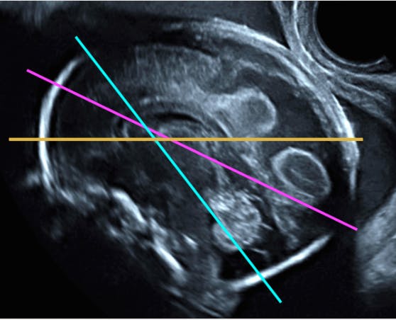
At 14 weeks, however, the clinician can see brain parenchyma and can visualize the brain stem, cerebellum, fourth ventricle, cortex, and vermis. A transvaginal ultrasound, when performed properly, can also visualize the corpus callosum as early as 14 weeks. Dr. Malinger notes that an ultrasound at this stage needs to be performed through the anterior fontanelle for best resolution.
Benefits of Late Second Trimester Ultrasound
A key point of Dr. Malinger's lecture was that a late second trimester or third trimester ultrasound is always indicated. He shared an example of when this can make a big difference in patient care. Dr. Malinger and his team published a letter to distinguish between choroid plexus cysts and ganglionic eminence (GE) cavitations. The first is a somewhat common occurrence that is not of major concern to the mother or physician. However, the latter condition has a very poor prognosis. Cysts often disappear during the late second trimester. However, images taken between 15 weeks and as late as 20 weeks demonstrate that what can appear to be cysts early on are actually GE cavitations. In this example, a late second trimester ultrasound helps clarify a diagnosis and determine appropriate course of action.
An ultrasound at 11 to 13 weeks allows for diagnosis of many abnormalities, but Dr. Malinger poses the question: Is this ultrasound the good one? Through research by his team and others, as well as a range of ultrasound images by experts in the field, Dr. Malinger demonstrated differences in performing ultrasound during the first versus second trimester. As the brain develops, a later trimester neuro-sonography allows the clinician to glean more information about the cranial structures, CNS, and brain formation. Waiting until 14 to 16 weeks can diagnose more abnormalities, allowing patients to make more informed decisions about their care.
Watch Dr. Malinger's video HERE.


