
Distinguishing different types of uterine abnormalities is challenging, particularly when using 2D ultrasound. However, sonographers increasingly use 3D ultrasound to more accurately diagnose mullerian anomalies, using the guidelines governing their area of practice.
While not all patients (even those with severe mullerian abnormalities) will have trouble conceiving or carrying a pregnancy to term, some, who have problems with fertility, miscarriage, and preterm delivery, require further investigation. A 2022 article in the Journal of Clinical Medicine refers to congenital uterine anomalies as "not uncommon," reportedly present in around 5% of the general population. However, the numbers are much higher in patients who have experienced multiple miscarriages or infertility.
Read on to learn about classifications of uterine anomalies, considerations for scanning, and frequent, and dangerous, diagnostic mistakes.
ESHRE Classification of Congenital Uterine Anomalies
In 2013, the European Society of Human Reproduction and Embryology (ESHRE) and the European Society for Gynaecological Endoscopy (ESGE) published a consensus paper classifying congenital uterine anomalies as follows:
- U0: Normal
- U1: Dysmorphic
- U2: Septate
- U3: Septate
- U3: Bicorporeal
- U4: Hemi
- U5: Aplastic
- U6: Unclassified
ASRM Mullerian Anomalies Classification 2021
In 2021, the American Society for Reproductive Medicine (ASRM) published a classification system for uterine anomalies, complete with diagrams and descriptive terminology, with the following categories:
- Müllerian agenesis
- Cervical agenesis
- Unicornuate uterus
- Uterus didelphys
- Bicornuate uterus
- Septate uterus
- Longitudinal vaginal septum
- Transverse vaginal septum
- Complex anomalies
Importantly, arcuate uterus was removed from the ESHRE classification system, while the ASRM considers this condition to be a variation of normal.
Scanning for Uterine Abnormalities
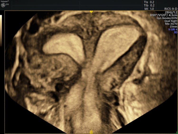
Significantly, using 2D imaging to diagnose mullerian anomalies tends to result in overestimating the anomaly or misdiagnosing it as the wrong type. In contrast, 3D transvaginal ultrasound can clearly visualize the uterus' shape, inside and out, without the need for hysteroscopy: it captures and displays images in three planes simultaneously. However, it is important to begin with a 2D scan, which will provide important information about the uterus (e.g., its size, position, and echotexture) as well as the endometrium and cervix.
After optimizing the 2D image, the sonographer should reduce the box size to display only the uterus, from fundus to cervix, and draw a curved line from one to the other. When the volume is obtained, the sonographer should optimize the images, by increasing contrast (if needed) and reducing artifacts. When the system has rendered the 3D image, it should automatically take measurements to provide a diagnosis. For a partial septate uterus, for example, these measurements will include interostial distance, septal depth, and the distance between the serosa and the interostial line.
Measuring from the fundus, an indentation of more than a centimeter (ten millimeters) indicates a bicornuate or unicornuate uterus. An indentation of seven millimeters or less is considered normal. The sonographer may also measure the length of the uterine cavity, as some patients, such as those with Turner syndrome, may have smaller uteri.
Diagnostic Mistakes are Costly for the Patient
Ultimately, a classification system exists so clinicians can assign risks to patients with uterine abnormalities. For example, researchers once associated the arcuate uterus with multiple miscarriages and preterm delivery. However, subsequent findings led to a dismissal of this relationship, according to the Jornal Brasileiro Reproducao Assistida.
One possible outcome of the clinical investigation of uterine abnormalities is surgical intervention. As such, an inaccurate diagnosis may expose a patient to an unnecessary, harmful surgical procedure. For example, a resection, while sometimes necessary, may also result in unintended uterine rupture; therefore, clinicians should approach resections with caution.
One anomaly which less-experienced sonographers over-diagnose is the bicornuate uterus, which may appear septate or even arcuate on 3D imaging. A smaller septum would represent a resectable case, and the 3D imaging would facilitate surgical planning as well as documentation. While the arcuate uterus is classified as normal in the United States, some practitioners may still flag it as an abnormality and inform the patient, creating confusion.
Even the most talented sonographers may easily generate distorted images by working in the wrong view. To get the best diagnostic images possible, working in the multiplanar view is recommended, rather than in the coronal plane or rendered view.
Providing an accurate and timely diagnosis for patients experiencing repeated pregnancy loss or infertility is often challenging. The diagnostic capability of 3D ultrasound can remove the uncertainty of an inaccurate diagnosis and ensure that uterine abnormalities are classified correctly according to ESHRE-ESGE or ASRM guidelines.
Interested in learning more? Click HERE.