The American College of Obstetricians and Gynecologists (ACOG) recommends the intrauterine device (IUD) as a first-line birth control method for nulliparous women. Still, this safe, highly effective contraceptive remains underused, with just under 12 percent of birth control users in the United States opting for either copper IUDs or hormone-releasing devices, according to ACOG.
New research suggests OB/GYNs can increase patient satisfaction with copper IUDs during insertion and beyond.
Realizing Contraceptive and Health Benefits
Sold under the trade name Paragard® in the United States, the copper IUD is clad in copper wiring, which interferes with sperm movement and implantation. Copper also intensifies a cytotoxic inflammatory response in the endometrium, but has no impact on ovulation. The devices may also offer non-contraceptive benefits, such as lessening the risk of cervical cancer and possibly reducing the likelihood of endometrial cancer.
Once implanted, copper IUDs are only slightly less effective than progestin-releasing IUDs, with a first-year failure rate of 0.52 versus 0.06 pregnancies per 100 women, reports a multinational study of more than 60,000 women.
Although Paragard® is FDA-approved for a decade of use, studies suggest the device may remain in place an additional two years without the risk of pregnancy.
Paragard® is right for most adolescents and women, per the device-maker, except for those with certain conditions, including an abnormally shaped uterus, unexplained bleeding, certain cancers, Wilson's disease, a pelvic infection or a recent infection of another type.
Answering Patients' Questions About Painful Insertion
IUD technology has come a long way since the early dangers of the Dalkon Shield. Today, the chief reason patients may shy away from IUDs is the fear of pain during insertion. Some pain is to be expected during the procedure, with women reporting it at speculum insertion, tenaculum placement, and IUD insertion, according to research published in the American Journal of Obstetrics and Gynecology. Whether the IUD was a hormonal or copper device appeared not to make a difference in patients' perceptions of pain.
Research tells us that a patient's perceived level of pain is influenced by their relationship with their physician, their anticipated level of pain, and their overall anxiety. Try to soothe patients' fears by describing what to expect, both before and during the procedure.
In terms of pain relief, timing the insertion of a copper IUD to coincide with your patient's period is not considered as helpful as it was previously, according to a systematic review published in Contraception. Instead, an application of lidocaine-prilocaine cream to the genital mucosa proved superior at alleviating pain when compared to methods including ibuprofen, misoprostol, lidocaine alone or a placebo, as reported in a meta-analysis published in Fertility and Sterility. Notably, most patients in the American Journal of Obstetrics and Gynecology study expressed a willingness to extend their appointment an additional 15 minutes so their doctor could apply numbing cream prior to device insertion.
Inserting the Copper IUD
Generally speaking, IUDs are inserted during a short exam using a speculum and a special insertion tool. Insertion involves passing the tube-like tool through the os and up the full length of the uterine cavity to the point of slight resistance, where the IUD reaches the top of the uterine fundus.
Copper IUDs are recommended for a uterine cavity of 6 cm to 9 cm, per the device-maker. Ultrasound imaging beforehand proves helpful for making sure the uterus meets size guidelines, thus ensuring a proper fit. A poor fit is a leading reason that nulliparous patients abandon this method.
Using Ultrasound in IUD Placement
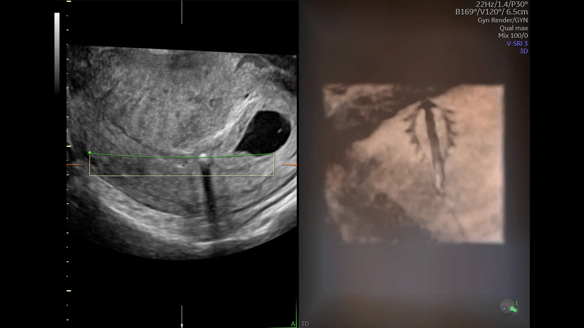
Ultrasound also proves beneficial in difficult IUD insertions, such as in women with cervical stenosis or a difficult cervical path. Such cases may require viewing the cervix or dilating before insertion. 3D ultrasound imaging can also identify distorted uterine anatomy, such as an anteverted or retroverted uterus or fibroids, that may affect a patient's contraceptive options.
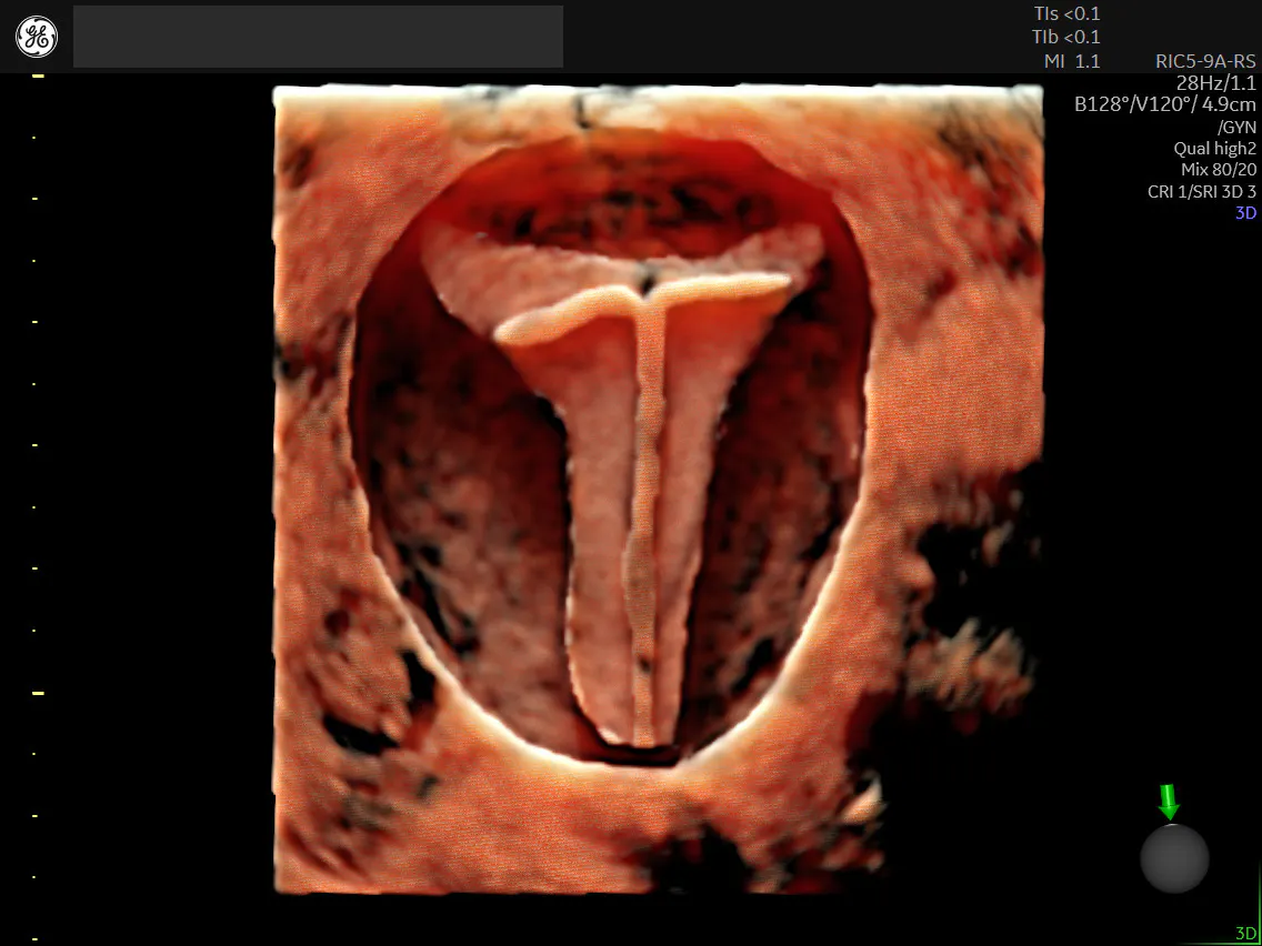
Uterus with IUD
Following insertion, a quick ultrasound is helpful to verify whether the IUD is in the correct position.
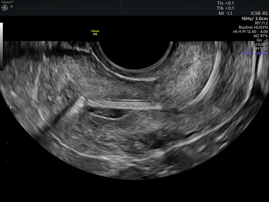
IUD with string
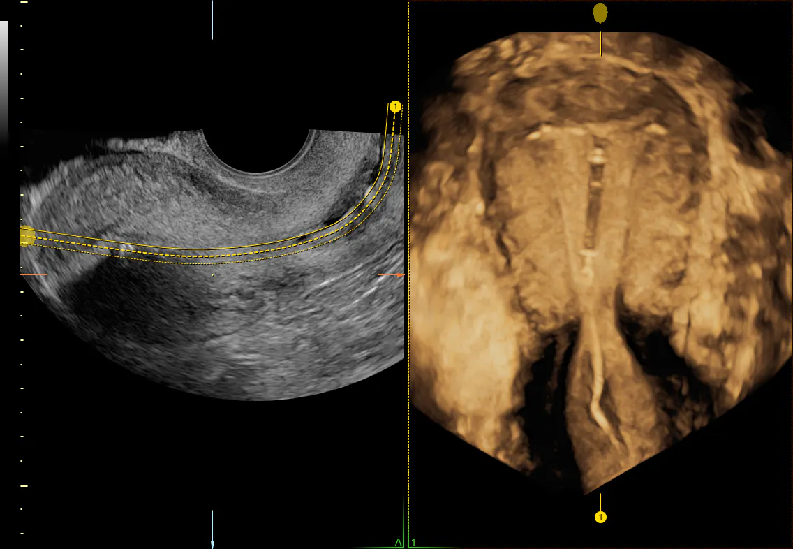
HDlive™ Studio rendered IUD
Aided with ultrasound technology, clinicians can feel confident that they are minimizing patient discomfort while doing the utmost to foster greater use of this convenient, highly effective birth control device.