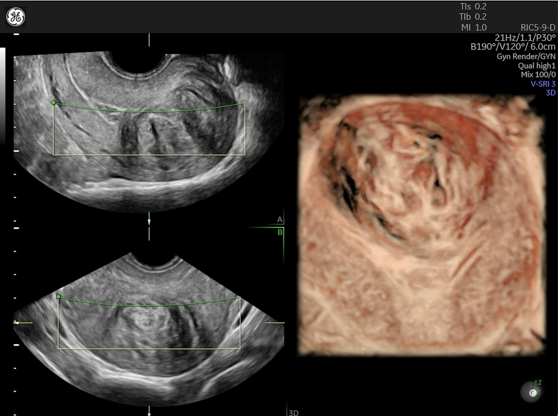
Recurrent implantation failure (RIF) is defined as three or more unsuccessful transfers of high-quality, genetically normal blastocysts. There are multiple reasons why an embryo might fail to implant, including uterine factors, tubal blockages or a difficult transfer process.
Once a patient reaches this point, is there a recurrent implantation failure treatment? What can reproductive endocrinologists recommend to frustrated individuals and families? First, physicians must identify the problem.
Uterine Factors: Anatomy, Polyps, Fibroids and Adhesions
When it comes to the uterus, there are several possible reasons for RIF. The patient might have a Müllerian anomaly, which is typically diagnosed during a saline sonogram or hysteroscopy. Of the possible Müllerian anomalies, only a septate uterus can be surgically corrected.
Other uterine issues are easier to identify and treat. Polyps — growths that attach to the inner wall of the uterus and can descend into the vagina — can range in size, with some as small as a few milli-meters and others as large as several centimeters. Although the connection between polyps and infertility is still poorly understood, research published in the Journal of Obstetrics and Gynecology of India found a higher prevalence of polyps in infertile and subfertile women.
Fibroids are classified according to their size and location in the uterus, following the International Federation of Gynecology and Obstetrics system or the updated PALM-COEIN system. Intramural fibroids, which grow within the muscular wall of the uterus, are the most common, but submucosal fibroids, which grow beneath the uterine lining and can crowd the uterus, often have the greatest impact on fertility. Fibroids can affect endometrial receptivity and prevent successful implantation.
The American Academy of Family Physicians recommends diagnosing fibroids using ultrasonography. Depending on the size and location of the fibroid, the physician may advise a myomectomy to improve future chances of implantation.
Adhesions, or scar tissue that involves the layers of the endometrial lining, the smooth muscle of the uterus (myometrium) or connective tissue within the uterus, are typically iatrogenic. For instance, they may form after a dilation and curettage. They may also form from retained products of conception. The AAGL and European Society of Gynaecological Endoscopy recommend treating adhesions, citing multiple studies showing improved pregnancy rates.
Endometrial Receptivity
Markers for endometrial receptivity are still debated in the literature, but endometrial thickness has long been used as a way to predict successful implantation. Endometrial thickness can easily be assessed using ultrasound. Research published in Fertility and Sterility reports that the uterine lining should be trilaminar in appearance. On ultrasound, this appears as an echogenic line in the middle surrounded by two thicker, darker hypoechoic lines and a well-defined hyperechoic outer wall.
If ultrasound reveals a thin endometrial lining, physicians may increase the patient's estrogen dosage or change the administration (patch, pill or injection). Additionally, because adequate blood flow is an important factor, reproductive endocrinologists may suggest more exercise, acupuncture or a medical approach, such as vaginal sildenafil. According to research published in the International Journal of Fertility and Sterility, vaginal sildenafil shows promise in aiding implantation for patients with multiple failed in vitro fertilization (IVF) or intracytoplasmic sperm injection attempts.
If ultrasound alone is not enough to assess the endometrium and recommend treatment, another option is an endometrial receptivity analysis. This ultrasound-guided uterine biopsy analyzes the appropriate number of hours the patient should be supplementing progesterone prior to a transfer. The results may indicate a recommended protocol change.
Additionally, research published in Frontiers in Endocrinology suggests that an intrauterine platelet-rich plasma (PRP) infusion can improve endometrial receptivity and reduce RIF. This new strategy involves centrifuging a small sample of the patient's own blood and reintroducing the plasma in the uterus. The growth factors and cytokines of the concentrated platelets promote cell growth and reduce inflammation in the endometrium. PRP infusion also has the advantage of using an autologous material, reducing adverse immune reactions.
Embryo Factors
Although RIF is defined as repeated failure to implant a healthy, high-quality embryo, the majority of embryos that fail to implant are genetically abnormal. Preimplantation genetic testing is not perfect; sometimes, it gives a false positive or false negative. Patients may also be uncertain how to proceed if their embryos are labelled as mosaic. Clinics should consider storing all embryos in the event the patient decides to retest abnormal embryos or transfer mosaic embryos (depending on the clinic or local rules for these transfers).
Other Causes of Repeated Implantation Failure
Inflammatory conditions, such as endometriosis and chronic endometritis, are known to interfere with fertility. Patients with endometriosis may take Lupron injections or Orilissa pills for several weeks or months prior to an embryo transfer. Treating chronic endometritis is typically as simple as a course of antibiotics.
Hydrosalpinx is when one or both fallopian tubes fill with fluid, which often spills into the uterine cavity. Tubal blockages may also be spotted on ultrasound with color Doppler. A systematic review and meta-analysis published in the Journal of Minimally Invasive Gynecology reaffirmed previous studies' conclusions that hydrosalpinx inhibits implantation and found that treating the condition, no matter which method is used, results in improved chances of pregnancy.
Preventing a Difficult Transfer
Sometimes, an embryo transfer is unexpectedly difficult. This may damage the embryo or cause the uterus to contract and reject the embryo altogether.
Physicians should perform a mock transfer prior to transferring another embryo. This is an opportunity to select the best catheter for the patient's body and ensure there are no anatomical reasons, such as scar tissue on the cervix, that might prevent smooth catheter insertion. When performing a mock transfer, identify the best place to put the embryo in the uterus, measure the length from the patient's cervix to that spot and map out the best route. Mock transfers can be performed about a month before a real embryo transfer.
Advising Patients
A failed IVF transfer devastates patients. They seek answers that a reproductive specialist cannot always provide. Physicians will want to thoroughly and compassionately explain the possible problems and solutions. Together, they may decide to move forward with one or more of the available treatment options prior to another transfer.


