
Ultrasound for fertility assessment and care is one of the most important tools that IVF physicians have at their disposal. From the initial consultation to the referral to an obstetrician, each step involves evaluation and treatment that is both facilitated and made less complicated by the use of ultrasound exams. Ultrasound is used to uncover the reasons for infertility, to help treat them and is also used to facilitate the IVF process itself.
This article recaps two lectures presented before the 2022 ISUOG World Congress by experts in IVF who use ultrasound to enhance patient care, and learn how their experience with both 2D and 3D ultrasound has shaped the way they approach IVF practice.
Dr. Sotirios Saravelos, Imperial College London
Dr. Saravelos has seen a shift from 2D to 3D and shares that he likes working with 3D ultrasound because it lets patients see their condition in real time and understand through visualization. He likes that the 3D modality also helps clinicians create models of complex anatomical anomalies to better understand how to plan treatment for their patients.
In the work-up phase prior to IVF treatment, there are any number of reasons for possible infertility, and the ultrasound provides the initial look into any potential problems.
Dr. Saravelos describes a patient who was previously diagnosed with a rudimentary obstructed uterine horn. She was prepared to have a surgical intervention but came to Dr. Saravelos for a scan. He diagnosed her with an accessory and cavitated uterine mass, which he had never seen before. However, after this first diagnosis, he began to see it more and more.
Polyps are more difficult to diagnose by 2D ultrasound, he says, and have been typically diagnosed by hysteroscopy. However, Dr. Saravelos states that by adding saline infusion during 3D ultrasound, intracavitary polyps can be visualized as plainly as during hysteroscopy. He recounts that there was no small struggle getting this observation published, even after reproducing this result in multiple patients, which shows how paradigms change reluctantly at times.
When to operate on fibroids is also a point of some debate. Dr. Saravelos says, however, that the location of the fibroid is key. He presents the case of a patient diagnosed with a unicornuate uterus on HSG (because there was no spill from the left tube), when in fact, a 3D ultrasound showed a fibroid blocking the internal os.
Adhesion bands may be a concern for patients who have had surgical management of pregnancy loss. Adhesions can be well-visualized with the addition of saline infusion during 3D ultrasound. Dr. Saravelos presents the case of a pregnant patient whose amniotic sac grew in a moon shape for an unknown reason, and the patient eventually lost the pregnancy. The patient was only diagnosed with an adhesion band after losing the pregnancy. Dr. Saravelos demonstrates that adhesion bands can be treated with ultrasound-guided balloon therapy in the office, without following up with a hysteroscopy.
When imaging the fallopian tubes, Dr. Saravelos suggests that using 3D inverse rendering will provide such detailed pictures that the sonographer will be able to visualize any mucous inside.
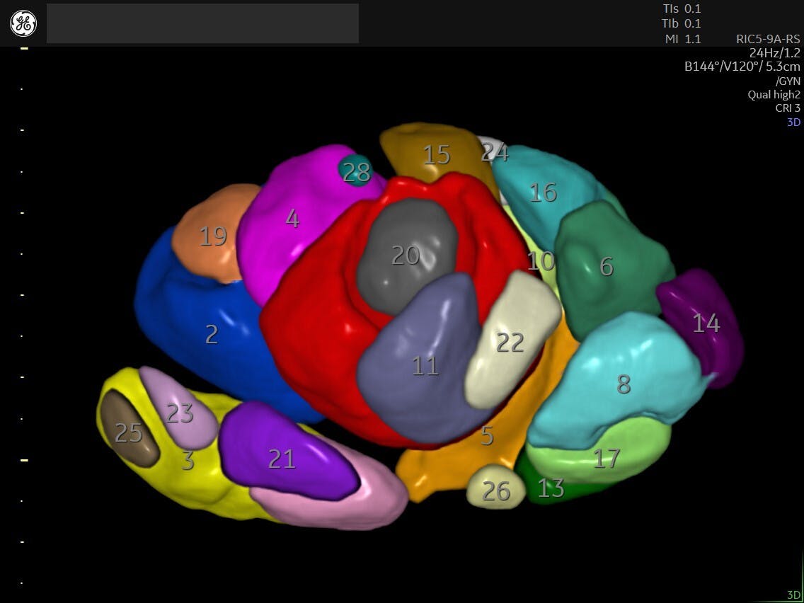
After ovarian stimulation, he relies on SonoAVC to aid with follicle tracking rather than counting by hand.
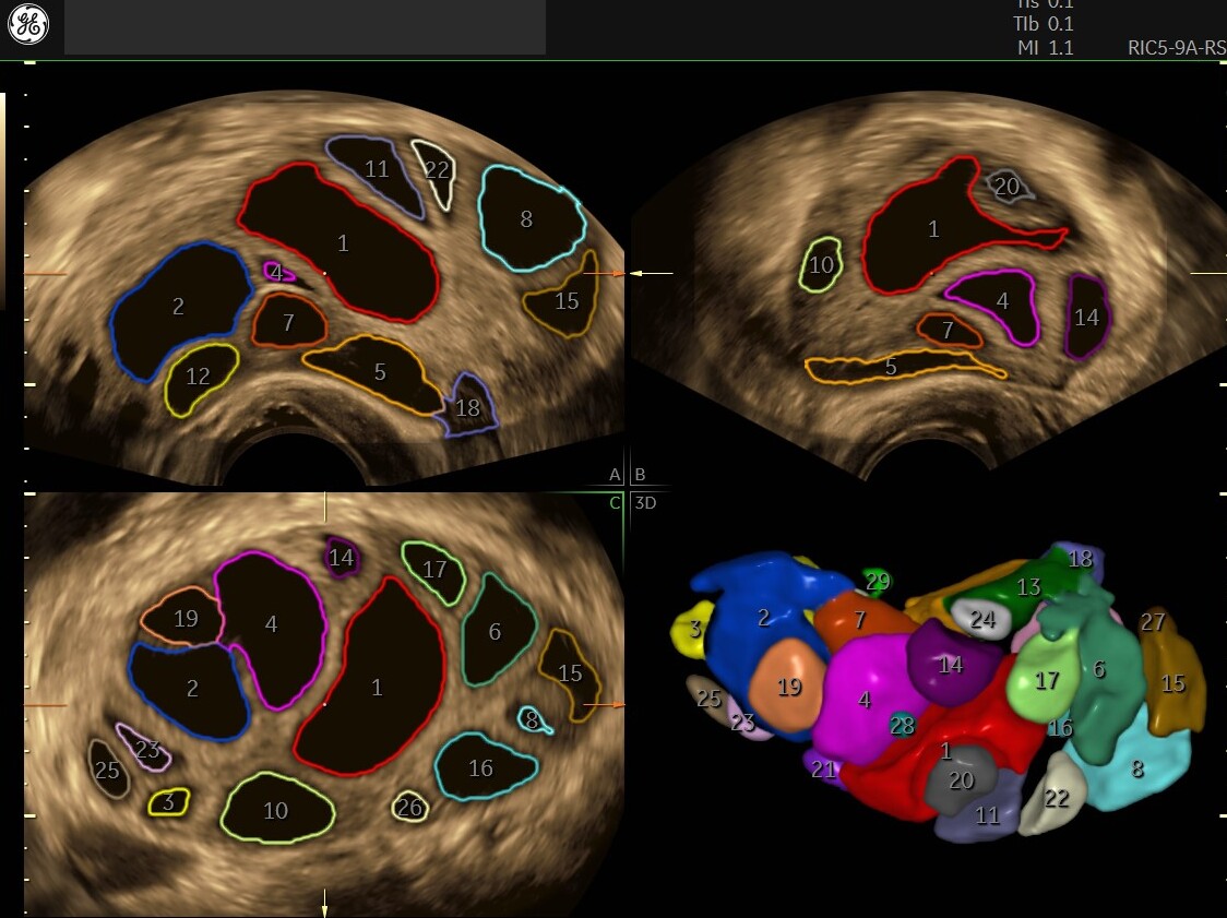
SonoAVC™ follicle automatically calculates follicle volume and dimensions
Dr. Saravelos has not found a clear clinical benefit of 2D versus 3D for the embryo transfer itself, but his practice has continued using 3D ultrasound for patient-centered reasons. First, the patient can see the embryo transfer more clearly. Second, if the embryo moves to the tube, for example, it is easy to refer back to the transfer images to demonstrate to the patient that the transfer was made properly.
Dr. Saravelos relays that he has been asked multiple times to perform 3D scans on pregnant patients with unclear diagnoses. For example, one patient had a single embryo transfer but a 2D scan diagnosed twins. However, Dr. Saravelos was invited to perform a 3D scan and found three fetuses.
What is apparent from this lecture is a shift from more invasive methods of imaging or even surgery to quicker, less invasive ultrasound imaging in fertility care. Even ultrasound professionals are seeing a shift to better methods, from 2D to 3D, and 3D methods are demonstrating that they can prevent surgical interventions in some cases.
Hear Dr. Saravelos explain how he uses 3D ultrasound
Dr. Simon Meagher, Monash IVF Group
The next expert, Dr. Simon Meagher, is affiliated with the Monash IVF Group, which produced the world's first IVF baby. He refers to ultrasound as a "one-stop fertility assessment." His lecture is extensive and full of useful pearls of wisdom, so we are just touching the highlights here.
Dr. Meagher prefers to see patients for fertility assessments during the second half of their cycle. He feels this is the best time to visualize the uterine cavity for mullerian anomalies. The downside of performing the scan at this time, he says, is that it may cause the sonographer to miss intercavity polyps, but performing a saline infusion during the exam can overcome this limitation.
In deciding whether to use 2D fertility assessment versus 3D, Dr. Meagher describes one of the most important features of 3D as being able to use the VCI Static feature to delineate tissue borders, for example, when trying to distinguish between endometriosis and a hemorrhagic cyst. Dr. Meagher displays corresponding 2D and Static 3D images of an endometrioma, pointing out the differences to demonstrate how much more clearly the VCI Static view shows the endometrioma.
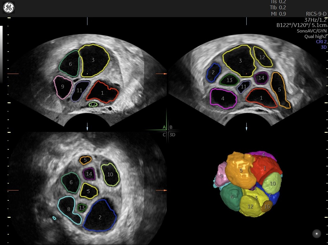
2D vs. 3D imaging with SonoAVC™
In evaluating the ovary during stimulation, Dr. Meagher doesn't find the VOCAL 3D rendering clinically useful, but instead stressed how helpful it is in facilitating the patient's understanding of what's happening in the ovary. Looking at the 2D image "means absolutely nothing to them," he says, but this underscores the physician's need for both 2D and 3D fertility assessment.
Again using the VOCAL tool to perform a volume assessment in the polycystic ovary to capture only the antral follicles, Dr. Meagher demonstrates the ability to image and automatically count 63 follicles in a patient, displaying follicles >6-7mm in red.
Interestingly, Dr. Meagher describes the precise evaluation of a "dermoid" cyst within a patient's ovary, using Static 3D technology to evaluate the different types of tissue present so that the patient would not assume that she needed to have the cyst surgically removed.
Dr. Meagher describes several useful incidental ultrasound findings in post-egg-collection patients such as soft-tissue masses, subcapsular hematomas and ovarian fibromas.
During the uterine ultrasound, Dr. Meagher suggests using a saline infusion of the uterine cavity to assess several conditions: asymmetry of the endometrium, cesarean scars and polyps that on 2D appear to be inside the uterine cavity.
When imaging for tubal patency, Dr. Meagher stresses that merely watching for flow through the tube to confirm tubal patency is not enough. He stresses that the sonographer needs to confirm "fill and spill" where the contrast agent fills the tube and spills out beside the ovary, since the tube may often be hidden behind the bowel.
One-Stop Fertility Assessment
While 2D is still a good and relevant imaging modality, 3D adds efficiency and precision to the ultrasound for fertility care. As both experts point out, 3D visuals help to engage the patient but tools like VOCAL, which aids in counting and measuring follicles, and SonoAVC™ follicle, which automatically calculates follicle volume and dimensions, give clinicians precise information to ensure that IVF care is being delivered on the right schedule. This information would have been previously calculated by hand or estimated by the visual 2D images. Static 3D can distinguish between tissue types, a feat previously only accomplished through surgical means.
Being able to provide precise, clear ultrasound for fertility care often steers patients away from unnecessary surgical procedures and speeds their journey on the pathway to achieving their ultimate goal.

Hear Prof Meagher explain how he uses 2D/3D ultrasound.