Measuring the cervical length is an essential obstetric ultrasound task for the primary purpose of predicting preterm birth (PTB), reports the International Society of Ultrasound in Obstetrics and Gynecology (ISUOG). PTB is defined as a delivery, regardless of the mode, before 37 weeks of gestational age. PTB is an adverse pregnancy outcome with significant implications on the neonate and family. Predicting PTB in a patient with a short cervix may lead to interventions that prolong the pregnancy to optimize outcomes, even allowing pregnancies to reach full term.
Having acknowledged the value of measuring cervical length, consider the following best practices regarding when and how.
When to Measure Cervical Length
Assessment is generally completed for screening purposes at mid-gestation, usually during the anatomy (morphology) ultrasound between 18 and 24 weeks. Although the Society of Maternal-Fetal Medicine (SMFM) recommends routine transvaginal screening for both high-risk (multiple pregnancies, those with a previous PTB, abnormalities of the uterus or previous cervical surgery) and low-risk patients, many centers continue to evaluate through transabdominal ultrasound as the first line. If an abnormality is identified on the transabdominal ultrasound, one may yield a more clear assessment of the cervix via transvaginal ultrasound. Very likely, the decision to perform only the transabdominal ultrasound is in part due to a combination of culture and convenience.
That said, there is some evidence published by the Nordic Federation of Societies of Obstetrics and Gynecology that the transabdominal ultrasound approach may be an effective screening step. Interestingly, despite the typical practices around the world, no national or international organization has endorsed a protocol for the measurement by transabdominal ultrasound, reports the Journal of Obstetrics and Gynaecology Canada.
On the other hand, there are several certification programs for cervical assessment that utilize transvaginal approaches, including FMF Certification and CLEAR. In addition, many reputable organizations — National Institute for Health and Care Excellence, Society of Obstetricians and Gynaecologists of Canada (SOGC), SMFM, and ISUOG — describe the transvaginal ultrasound approach as the gold standard.
How to Accurately Measure Cervical Length
According to the latest guideline published by the SOGC (Guideline No. 401), clinicians should consider the following instructions and best practices regarding transvaginal ultrasound:
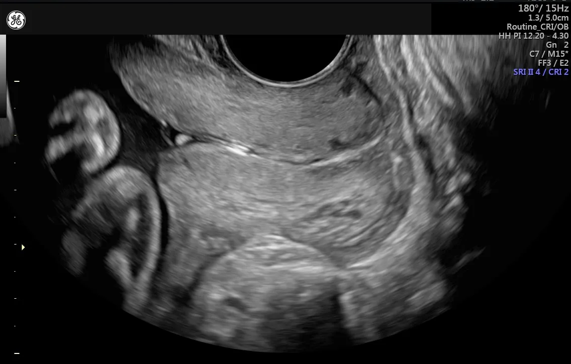
- The cervix should occupy a large portion of the image (~75 percent).
- The following landmarks should be identified:
- The anterior and posterior cervical thickness should be similar.
- Calipers should be placed where the anterior and posterior cervical walls touch the internal and external cervical os and should not extend beyond these points.
- A minimum of three measurements are taken over three minutes, and the shortest image should be used to determine the cervical length.
- A straight line or using a segmental approach can be used depending on the curved nature of the cervix.
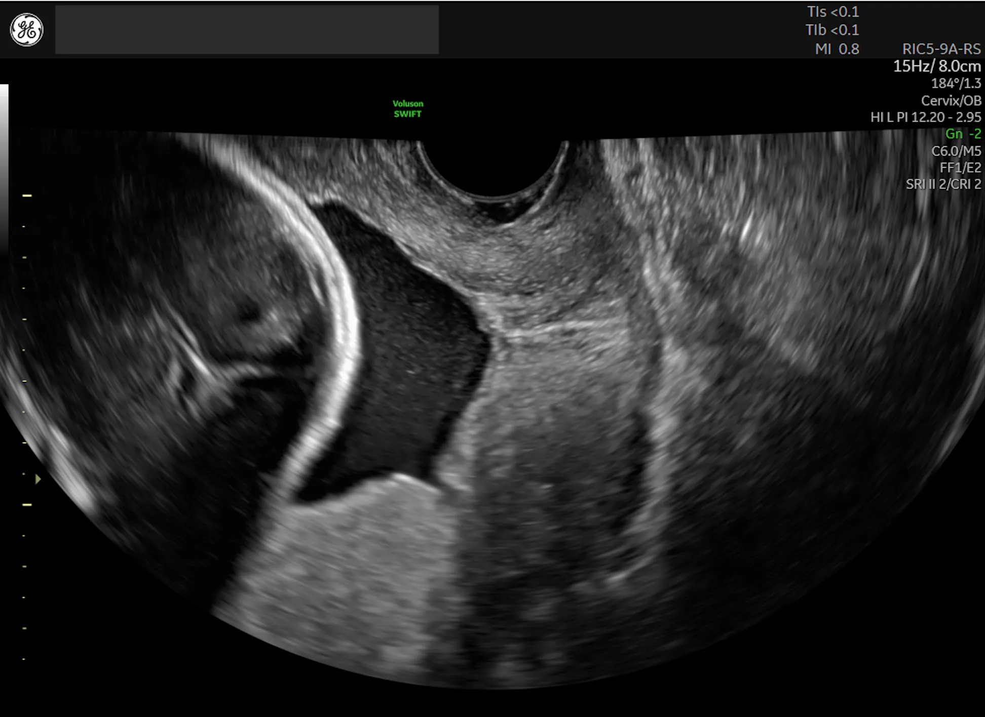
- The presence or absence of funneling, defined as the protrusion of the amniotic membranes into the cervical canal, should be noted.
Unique requirements
For the transabdominal ultrasound approach:
- Ensure the patient is in a supine position, with as close to 0 degrees as possible.
- A full bladder is not required, but some urine in the bladder may optimize the chance of acquiring an adequate image, says a study in the Journal of Ultrasound in Medicine.
For the transvaginal ultrasound approach:
- Ensure the patient is in a dorsolithotomy position.
- Ensure the patient has an empty or near-empty bladder.
- The right balance of pressure and image quality needs to be achieved. This can be facilitated by identifying the cervix from the anterior vaginal fornix, then withdrawing the probe until the image blurs to minimize compression on the cervix by the probe. The probe should then be reapplied just far enough to obtain the best image. Note: Excess pressure may artificially lengthen the cervix and funneling will be obscured.
- Suprapubic or fundal pressure may be applied for approximately 15 seconds to watch for cervical change, including funneling. Note: Exhibit caution in applying further pressure if there is a noticeable short or open cervix in the absence of pressure.
The Value of First-Trimester Measurements
According to an article published in BJOG: An International Journal of Obstetrics & Gynaecology, some argue there may be value in performing an assessment earlier in the gestation during the 11+0 to 13+6 week scan for the same purpose of evaluating the risk of preterm birth. In combination with clinical history, cervical length at the 11–13+6 week interval may detect roughly half of pregnancies delivered before 34 weeks gestational age, with only a 10 percent false positive rate. Although first-trimester screening for cervical insufficiency is not yet adopted or integrated into national or international guidelines, this may be a relevant approach in the future.
Assessment of the cervix via transvaginal ultrasound in mid-gestation for shortening and/or funneling is the most reliable method to predict PTB and attempt to positively influence the remainder of the pregnancy, optimizing obstetric and neonatal outcomes.