
Prenatal ultrasound has made vast advances in the past few decades. Not only can obstetricians safely perform ultrasounds up until the time of birth to monitor a fetus's health, but they can also diagnose some fetal conditions in the first trimester. One of the most important tools in monitoring fetal health is color Doppler.
Color Doppler and Safety
Color Doppler measures movement and displays it in color. It is used in a complementary manner with spectral Doppler. Color flow is used to give direction and determine high and low velocity. It can detect turbulence, display a color flow map and give an overview of flow within the imaged region. Spectral Doppler instead zeros in on a specific site that needs further examination, allowing the operator to calculate velocity and indices and examine the flow waveform.
Although prenatal ultrasound is considered to be safe, it can have adverse effects when used improperly. The American Institute of Ultrasound in Medicine (AIUM) recommends that color Doppler should not be used routinely but instead when clinically indicated, such as when evaluating for risks of trisomies or evaluating the placenta.
AIUM recommends that spectral Doppler be used with caution during the first trimester because of the inherent risks to the fetus. It can, in theory, be safely used to view the maternal uterine arteries as long as the fetus is not within the ultrasound beam. During the 11-14 week exam, AIUM urges obstetricians to only use spectral Doppler for "specific clinical indications."
Ultrasound is known to cause tissue heating, cavitation (the formation of gas bubbles), and streaming in fluids and tissues. Tissue heating in the first trimester is associated with teratogenic effects. The U.S. Food & Drug Administration (FDA) has addressed this issue by regulating the spatial peak time-averaged intensity (I-SPTA), which is associated with temperature increase more than other measures. The FDA limits the I-SPTA to 720 mW/cm 2 while placing "more responsibility on the user to understand the output measurements and to use them in their scanning." To reduce the effects of streaming and cavitation, the FDA regulates the mechanical index at a maximum of 1.9 (with an exception for ophthalmic scanning).
To this end, AIUM abides by the ALARA (as low as reasonably achievable) principle and suggests using the ultrasound machine's built-in settings after reviewing them to ensure they are appropriately set. The power should always be set at the lowest level to provide the images. The user should:
- Be familiar with the limits of the mechanical and thermal indices.
- Avoid having the transducer in a stationary position to avoid dwell time.
- Avoid eyes and bone.
- Keep the scanning time to a minimum.
Using Color Doppler as a Prenatal Diagnostic Method
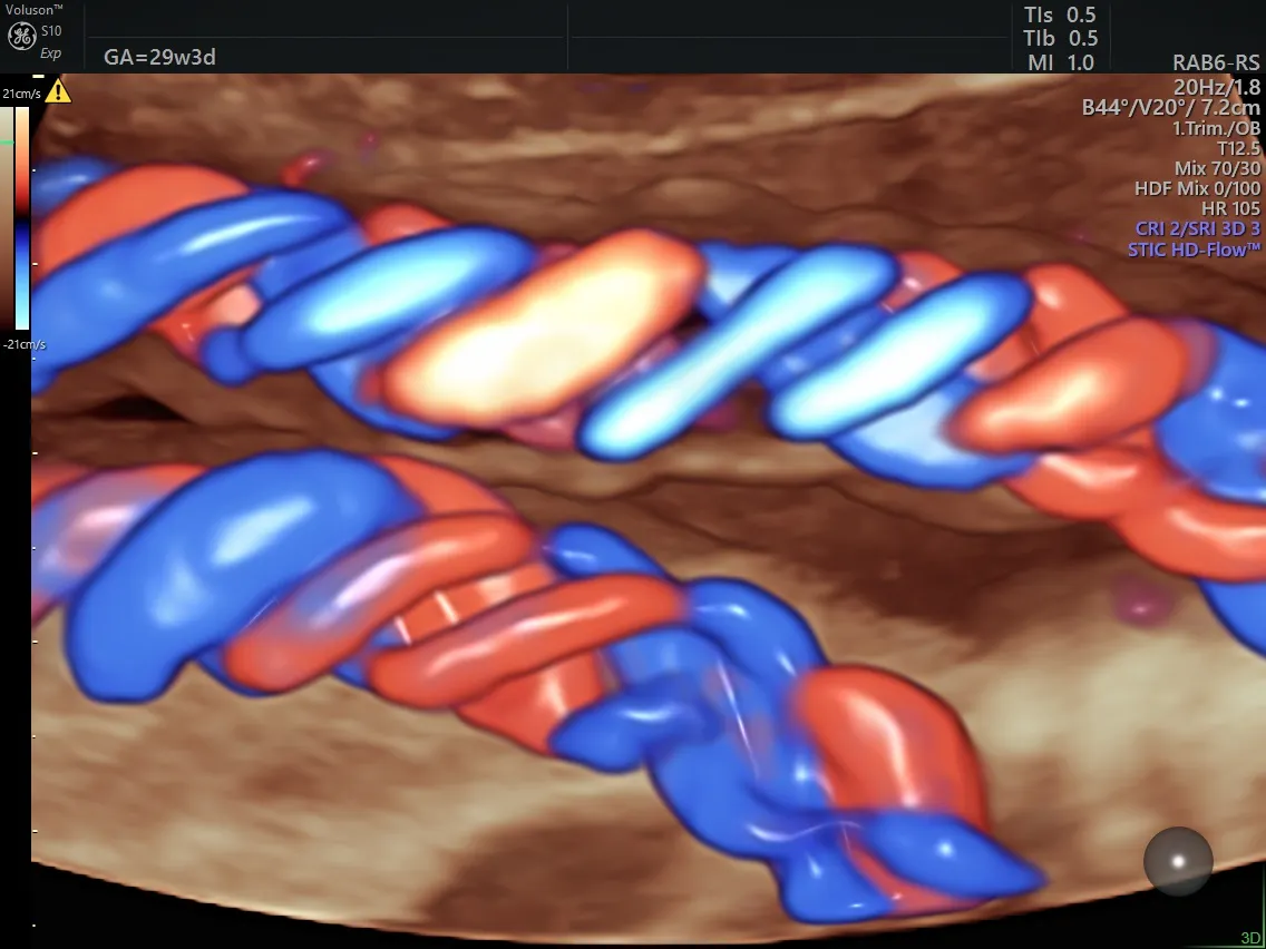
Generally, in the color Doppler image, red indicates blood or fluid that is flowing toward the transducer, and blue is flowing away from the transducer. The color saturation indicates velocity, with lighter shades of red or blue indicating higher velocities.
One can see the utility of color Doppler in detecting fetal heart abnormalities. While the heart's anatomy may not be apparent on 2D ultrasound, color Doppler can reveal the flow patterns. Heart defects such as tricuspid regurgitation or shunting ventricular septal defects — which may be associated with syndromic conditions —can be seen with color Doppler but not with gray-scale mode alone.
Ultrasound in Obstetrics and Gynecology refers to color Doppler as "essential" in evaluating fetuses with known congenital heart disease (CHD). In addition, the detection of CHD can be improved by using color Doppler and further enhanced with 4D ultrasound.
Fetal Medicine notes that color Doppler is useful in evaluating not only the fetal vessels of the heart, placenta, umbilical cord, kidneys and intracranial vessels but also in investigating fetal tumors and resolving a differential diagnosis of oligohydramnios.
Radiopaedia states that flow within the umbilical vein should be non-pulsatile (after around 13 weeks). Pulsatility in this vein may indicate chromosomal abnormalities and may be associated with increased perinatal mortality. However, these findings should be approached with caution, since fetal hiccups or even the movement of the fetus's breathing can cause these irregularities. Flow abnormalities within the umbilical artery can signify placental insufficiency. Preeclampsia should be suspected upon this finding. Intrauterine growth restriction can also result from placental insufficiency.
Other conditions involving vessels of the placenta and umbilical cord that can be identified with color Doppler include:
- Placenta accreta (the placenta attaches too deeply to the uterine wall)
- Placenta previa (the placenta covers the cervix)
- False versus true knots of the umbilical cord
- Vasa previa (the fetal vessels inside the membranes are close to or across the cervix)
The middle cerebral artery exam can indicate fetal cardiovascular distress, anemia or hypoxia, intrauterine growth restriction, twin-to-twin transfusion syndrome and twin anemia polycythemia sequence. In examining the middle cerebral artery, color Doppler helps identify the cerebral vessels. An increased blood supply to the brain can indicate fetal hypoxemia since the fetal system decreases blood to other organs and lower regions of the body in an attempt to spare the brain.
Color Doppler obstetric ultrasound is a versatile tool that can enhance investigations into both maternal and fetal conditions. When performed by a skilled and knowledgeable clinician, this prenatal ultrasound tool can uncover maternal and fetal conditions that might otherwise remain hidden. Used properly, this is a safe and noninvasive method of diagnosing conditions of potential concern.