

The grayscale images produced by 2D and 3D ultrasound provide excellent morphological assessment of adnexal masses. Color Doppler complements this ability by allowing visualization of blood flow within a mass. Using ultrasound with both grayscale and color Doppler can help clinicians detect, diagnose and manage adnexal masses, and help their patients with treatment options in a timely manner.
Predicting the Risk of an Adnexal Mass
It's possible to estimate with a high degree of certainty whether or not an adnexal mass is malignant. Recognizing certain ultrasound markers and determining whether a mass is vascularized can help a gynecologist decide whether a particular patient needs conservative management, surgery or an oncologist.
The International Ovarian Tumour Analysis (IOTA) Group created a few of the most widely accepted predictive models: the IOTA Simple Rules and the ADNEX risk models. The Simple Rules model is a " preoperative classification system for ovarian tumors", while the ADNEX model "estimates the probability that an adnexal tumor is benign, borderline, stage I cancer, stage II-IV cancer or secondary metastatic cancer (i.e. metastasis of non-adnexal cancer to the ovary)."
The IOTA Simple Rules model consists of 10 features, five of which are considered benign, or B-features, and five of which are considered typical for malignant tumors, or M-features. Besides accounting for the presence or absence of ascites and acoustic shadows, the size and number or papillary projections and other mass characteristics seen on ultrasound, this model also includes a color score. The absence of color Doppler flow is assigned a color score of 1, and is considered benign (B-feature). The color score increases with the amount of color flow seen, up to a color score of 4 (very strong blood flow), which can indicate a malignant or M-feature.
Many tumors may be classified by using the IOTA Simple Rules model. If at the end of the examination, the classification is considered to be "inconclusive" according to the model, then further examination, such as with the ADNEX model, is needed.
The ADNEX model uses nine variables, six of which are taken from ultrasound examinations. The ultrasound variables include diameter of the lesion and its largest solid component, whether more than 10 locules are visible, the number or presence of papillary projections and the presence of acoustic shadows and ascites. The other three variables are clinical in nature, and include patient age, serum CA-125 level and the type of center (oncology referral center or other) where the exam is performed.
Color Doppler: How It Works and Why It Helps
While 2D and 3D ultrasound alone are designed to visualize structures or tissues inside the body, the inclusion of color Doppler imaging can reveal important clinical clues by showing the presence or absence of blood flow, as well as the direction of blood flow. The International Journal of Oncology notes that many ovarian pathologies have clear distinguishing markers on ultrasound. For example, cyst contents will appear anechoic but will have no vascularization, while a teratoma, usually a benign mass, will have a mixed echogenicity because of its contents. A teratoma can also be vascularized if it is malignant.
With experience, ultrasound operators can easily distinguish between normal and increased vascularity in the adnexa, and recognize when an increase might signal an inflammatory process rather than a malignancy. Practictioners my utilize protocols built into their ultrasound systems, such as Scan Assistant, which guides the user through ultrasound parameters like the ADNEX risk model to ensure that screening is consistent, efficient and reproducible.
Most ovarian masses are benign, but considering the high mortality of ovarian cancer, adding color Doppler to a scan can add another element of certainty to the diagnostic process. Early detection and intervention can help improve a patient's chances at any age and risk level.
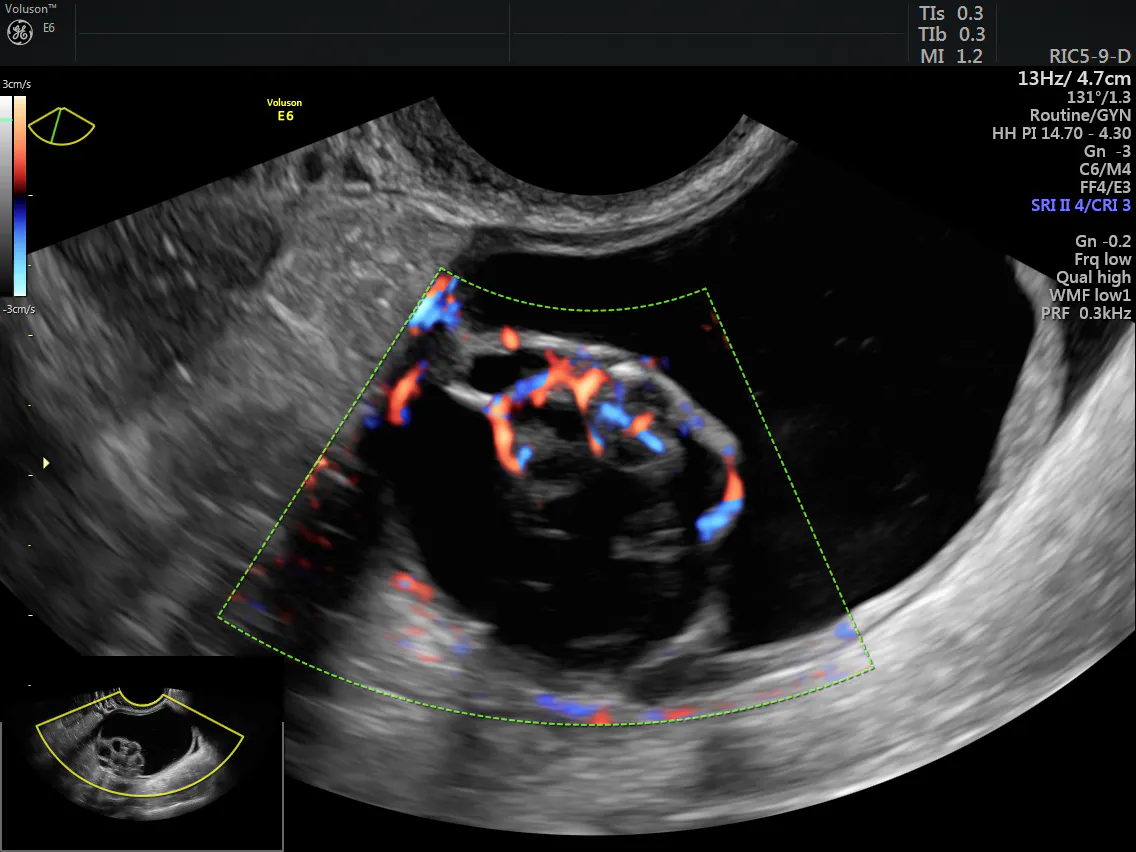
Ovarian mass with color flow


