More women than ever are delaying parenthood to pursue an advanced education or further their careers. Many women expect pregnancy to come easily later in life, but they might be unpleasantly surprised by the rate at which their fertility declines. A woman is most fertile before age 30; once women reach their mid 30s, fertility drops rapidly, according to the American College of Obstetricians and Gynecologists. By age 45, getting pregnant without medical help is unlikely.
Ovarian reserve testing is becoming increasingly popular as more women choose to have children later and doctors attempt to predict their likelihood of achieving pregnancy. This evaluation uses a combination of blood tests and ultrasound to provide information about diminished ovarian reserve (DOR) and can inform about the use of assisted reproductive technology (ART).
Your patients likely have questions regarding ovarian reserve testing. Sharing information about the accuracy of this test and the steps involved will help provide them with the best possible care.
Ovarian Reserve Testing Indications
The American Journal of Obstetrics and Gynecology notes that ovarian reserve testing is indicated in several different situations, including:
- Before undergoing infertility evaluation or treatment.
- For planning individual ART ovarian stimulation protocols and dosing.
- A history of early menopause or premature ovarian failure.
- Polycystic ovarian syndrome (PCOS).
- Fertility preservation before or after treatment with gonadotoxic agents.
- Preparation for ovarian surgery in women of reproductive age.
- Diagnosis and recurrence surveillance for granulosa cell tumors.
- Perimenopause.
- A BRCA-1 or FMR1 premutation.
Additionally, women considering elective egg freezing or oocyte donation should undergo this screening.
Beginning With Blood Tests
Ovarian reserve testing begins with a blood test to measure levels of follicle stimulating hormone (FSH), a hormone associated with ovarian aging. Generally, these tests are performed on the second or third day of a woman's menstrual cycle. According to the University of Colorado, elevated FSH levels on day two or three of the menstrual cycle correlate with reduced ovarian reserve.
Clinicians also measure estradiol to help evaluate ovarian function. Typically, estradiol levels fluctuate over the course of the menstrual cycle. Elevated levels on day three of the cycle may also indicate dwindling ovarian reserve.
One critical hormonal measurement used to estimate ovarian reserve is the anti-Mullerian hormone (AMH) test. Many clinicians consider AMH an important parameter for assessing ovarian reserve (in conjunction with other parameters such as the antral follicle count) since levels tend to remain constant for perimenopausal women, according to Menopause Review. Age-specific AMH values have been identified through various research studies. It is generally accepted that AMH values of 1ng/ml or less represent diminished ovarian reserve.
Ultrasound's Role in Ovarian Reserve Testing
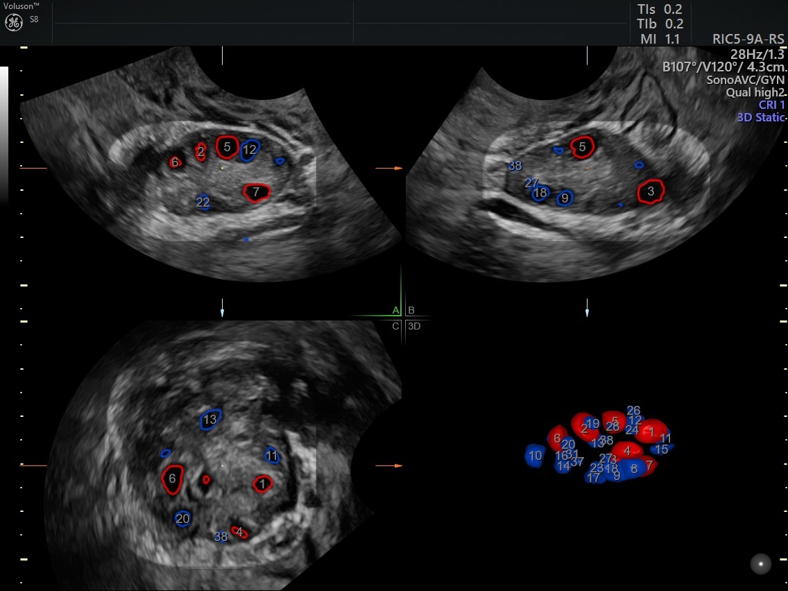
In addition to hormone measurement through blood tests, ultrasound is used to count small (antral) follicles in the ovaries. Technologies like the SonoAVCantral tool allow users to determine the number of follicles present that are also within pre-specified size ranges (i.e. 2-10 mm). The SonoAVCantral tool may be particularly useful for patients with a high antral follicle count, allowing clinicians to visualize and interpret the count more efficiently.

Follicles of the same size range are shown with the same color and are counted automatically with SonoAVCantral.
Ovarian reserve test results are not an absolute indicator of fertility problems, and you should be careful to explain to patients that ovarian reserve deficiencies do not necessarily mean complete infertility. However, it is important to be honest about test results and the likelihood of achieving viable pregnancy, even with the use of ARTs such as in vitro fertilization. Be sure to explain the purpose and process of ovarian reserve testing to your patients, including information about how you will plan further fertility treatment based on the results.


